FTC disclaimer: This post may contains affiliate links and we will be compensated if you click on a link and make a purchase.
An electrocardiogram (ECG or EKG) is a procedure used to obtain a record of the heart’s electrical activity. The test can help diagnose and/or monitor heart problems. Results from an ECG can also be used to determine if treatment is necessary.
Electrocardiogram (ECG or EKG) is a test used by a health professional to assess the heart’s electrical activity from the body’s surface.
This is a very reliable test. This test can be done at rest or with stress (on a treadmill).
What is an electrocardiogram?
An ECG is an electrocardiogram, a test that measures the heart’s electrical impulses over a specific period. E, C, and G stand for Electro-Cardio-Gram. Below is an explanation of the electrocardiogram.
It is a highly sophisticated way of learning how your heart functions and the best way to pinpoint what is wrong. Your doctor may refer you to a cardiologist who will read the rhythm strip and suggest a course of medical care.
ECGs have helped millions of people avoid major heart problems. It is a well-used tool that helps you and your doctor make educated decisions about your healthcare. If your doctor orders an ECG for you, take it seriously and find out what is going on with your heart.
Signs That You May Need An Electrocardiogram (EKG)
An EKG is beneficial in many heart conditions like;
- angina (chest pain)
- myocardial infarction or heart attack
- high blood pressure or hypertension
- heart failure
- hyperkalemia (high potassium)
- pericarditis
- hypothermia
The purpose of Electrocardiogram
Health care providers use an electrocardiogram for many purposes. These include assessing;
- the heart rate
- heart rhythm abnormalities
- conduction abnormalities
- any chamber enlargement
- signs of heart attack
- any electrolyte abnormalities
The risk with Electrocardiogram
Some potential risks that may come with having an ECG are:
- Electrode placement
- Incorrect electrode placement
- Noise levels
- Radiation.
It is essential to be aware of these risks to control them during the procedure.
What is recorded during the ECG test?
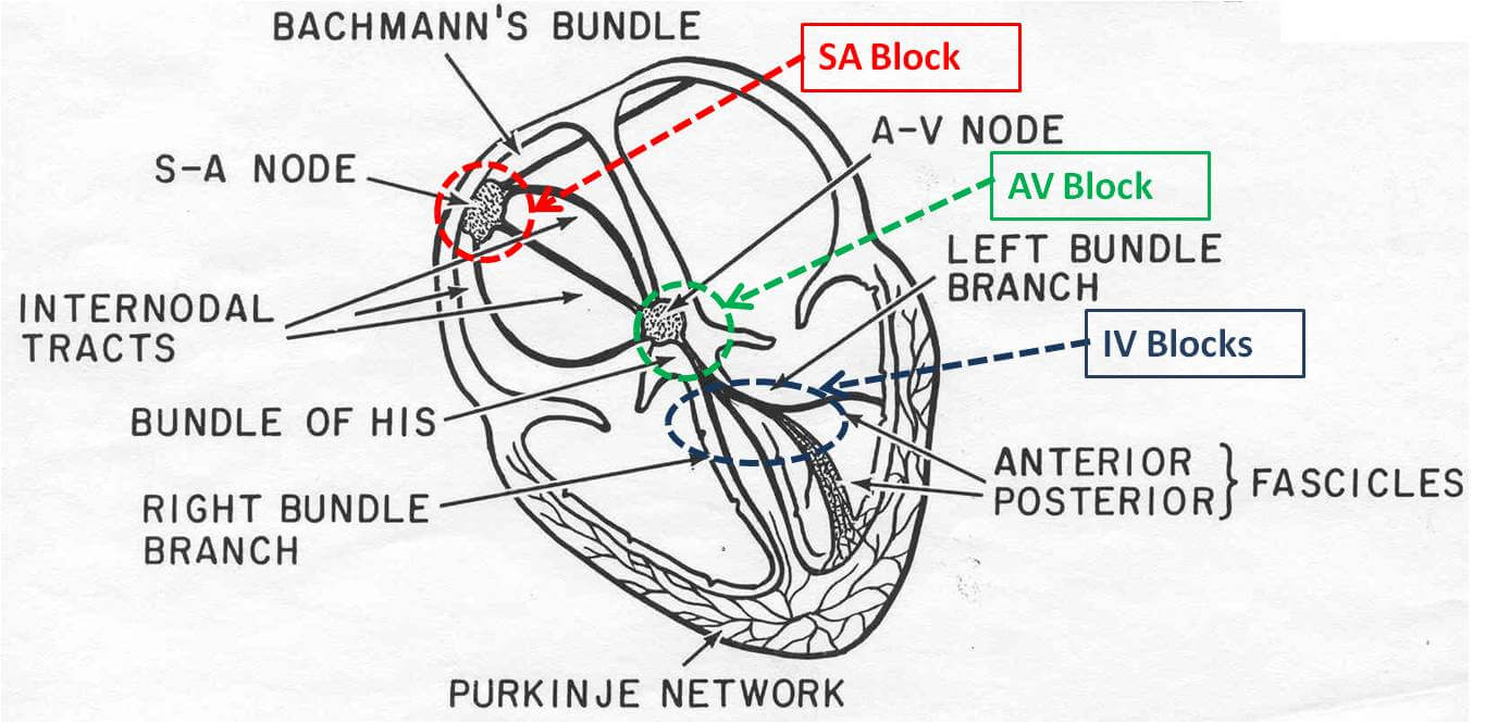
The human heart is composed of four chambers. The upper two are collectively called atria which are named according to their location in the human body.
Thus the atrium on the left side is designated as the left atrium (LA), and on the right, the right atrium (RA).
The lower half chambers of the heart are called ventricles, also named according to their location.
Thus, these four chambers comprise the human heart and function to pump blood throughout the body.
Each chamber has a distinctive role in circulating blood. The atria receive blood from the circulation, whereas the ventricles pump blood into the circulation.
Blood circulation sequence:
- Right Atrium via Superior and Inferior Vena Cava receives deoxygenated blood from the body’s tissues
- The blood passes through the tricuspid valve; valves are placed between chambers to prevent the backflow of blood
- The right ventricle pumps blood to the pulmonary artery passing through the pulmonary valve
- The blood travels until it reaches the lungs for its oxygenation
- Oxygenated blood is carried back to the Left Atrium via pulmonary veins
- The Left Atrium contracts and pushes it through the mitral valve, allowing it to flow to the left ventricle with less force.
- Blood passes through the aortic valve from the left ventricle and flows into the aorta for distribution to the rest of the body.
The human heart is so unique that it has its own blood supply, a vital feature knowing that the heart muscle never rests; it works continuously even as we sleep.
These coronary arteries supply oxygen to both the heart’s muscles as well as its nerves.
The heart also possesses its own electrical system via the nerves. Electrical current runs through these nerves to initiate contraction, and these electrical currents can be recorded and traced with an ECG test.
Types of electrocardiograms
Resting electrocardiograms
The most common type of electrocardiogram is a resting electrocardiogram or ECG. Resting electrocardiograms earn their name because this type of electrocardiogram is performed while the patient is lying down.
Resting Electrocardiogram is often part of a routine physical exam.
How effective is the resting electrocardiogram?
The answer is not very effective. Usually, the resting electrocardiogram will not reveal anything unless your heart has major damage.
Exercise electrocardiograms or Stress electrocardiograms
Exercise electrocardiograms are done when the patient exercises, such as walking or running on a treadmill with the electrodes attached to the chest.
Moreover, Exercise electrocardiograms are also called stress electrocardiograms because it tests the patient’s heart during the stress of physical activity.
Also, Exercise electrocardiograms or stress electrocardiograms often reveal heart damage caused by narrowing arteries, restricting the amount of oxygen getting to the heart muscle.
Electrocardiograms or ECGs are sometimes referred to as the Treadmill test.
Other tests similar to electrocardiograms
Some other tests include:
- the Thallium test
- Doppler ultrasonography test
- the MRI (Magnetic resonance imaging)
- the Echocardiogram, and
- Chest X-ray
Understanding How Your ECG Is Interpreted
Occasionally your doctor will order complicated ECG tests for you so that they can determine what is currently ailing you. If you have a heart problem, they may order an ECG.
Your health care provider will ask you to lie on a bed or sit comfortably.
The doctor will place electrodes on certain areas of your body to help measure how well your heart is working.
Electrocardiograms attach electrodes to the patient’s chest, wrists, and ankles. The electrodes measure electrical signals and record them on a paper or computer screen.
Twelve electrodes are attached to your body to measure your heartbeats. These electrodes are called leads and help the doctor ‘see’ your heart’s electrical activity from various angles.
The ECG will show them what part of the heart is affected by the condition you are suffering from and which way the depolarization of your heart is traveling.
These leads measure the cardiac axis of your heart, and each lead measures something different so that it makes a wave on the tape showing how your heart is working.
Also, the leads attached to your body record a different perspective of your heart’s electrical workings.
What ECG leads or electrodes measure?
The leads corresponding to a different spot on the heart can help identify things such as heart injury or other acute coronary conditions.
Physicians analyze any abnormalities in the pattern of the waves to identify possible signs of heart problems or heart disease.
If the leads measure areas of the heart next to each other, they are called contiguous. This is done to help see if there is a heart problem or something that is a one-time incident.
Some leads measure the heart’s ventricles and the heart’s surface.
If the doctor needs to look for a specific signal from your heart, they can program the ECG machine for that particular electrical pulse.
Further, it can monitor regular cardiac rhythm as well as any inconsistencies. This helps pinpoint where low signals originate from.
Once your doctor has performed the ECG, he will read the results from a ‘rhythm strip‘ that prints out a graph of your heart rhythm.
Each of the twelve leads is represented on the strip. The strip also contains a time stamp, and the graph shows the doctor exactly when in the ECG the reading occurred. It is not unusual for the heart’s rhythm to change based on what the leads report.
If you have heart problems or think you are having a heart problem, contact your physician immediately. They may order an ECG to find out what is going on.
Deciphering ECG Test
An Ecg Test is one of the many diagnostic tools used to measure and record a human heart’s electrical activity when the heart muscle’s cells in both the atria and the ventricles contract. This is useful in deriving diagnoses once the results are ready for interpretation.
The results of the Ecg Test will appear on a roll of paper as a series of waves and frequencies on which physicians base their interpretations.
The following are waves that one normally sees in a typical electrocardiogram result, but these waves vary depending on the heart condition of a patient.
The electrical voltages generated by the heart through Ecg Test are recorded as P, QRS, and T waves. The causes for these waves are as follows.
P Wave
P-wave is caused by the spread of depolarization over the left atria; the atria contracts right after. This contraction causes a minimal rise in the atrial pressure curve right after the P wave.
QRS wave
From the start of the P-wave, 0.16 seconds later, QRS waves appear resulting from the depolarization of the ventricles, which causes the ventricles to start their contraction, followed by a rise in the ventricular pressure curve. The QRS complex begins a little before the start of ventricular contraction.
T-Wave
The ventricular T-wave depicts the stage of repolarization of the ventricles wherein the muscles of the ventricles begin to relax. T waves appear before ventricular contraction ends.
How to use the ECG results?
Heart rate
The normal heart rate is between 60-100. This is a very reliable test to show if the heart is beating fast (tachycardia) >100 or beating too slow <60 (bradycardia).
Heart rhythm
Normally the heart pumps regularly (normal sinus rhythm). The ECG can pick up any irregular heartbeats and abnormal waves.
Conduction abnormalities
ECG can detect any conduction abnormalities. The duration of the electrical activity will also be detected.
Conditions like these include;
- Right and left bundle branch block
- Fascicular blocks
Chamber enlargement
Conditions like high blood pressure can cause the walls of the heart (ventricles) to thicken. This is known as hypertrophy. An EKG can detect any hypertrophy or enlargement present.
Heart attack
In angina, heart attack, or myocardial infarction, an ECG will show any abnormalities. The doctor uses these results to assess the extent of these conditions. These abnormalities could be;
- ST-segment elevation
- T wave invasion
These are the most popular uses of an ECG.
The Bottom Line
The electrocardiogram procedure, test, and results are valuable tools that can help a healthcare professional diagnose and treat a patient.
When used together, it can provide a comprehensive view of the patient’s cardiac health. Healthcare professionals should know how to use these tools and understand the results they produce.
